Miiling invisible structures
Geomill326’s digital imaging allows milling along otherwise invisible growth lines and striations
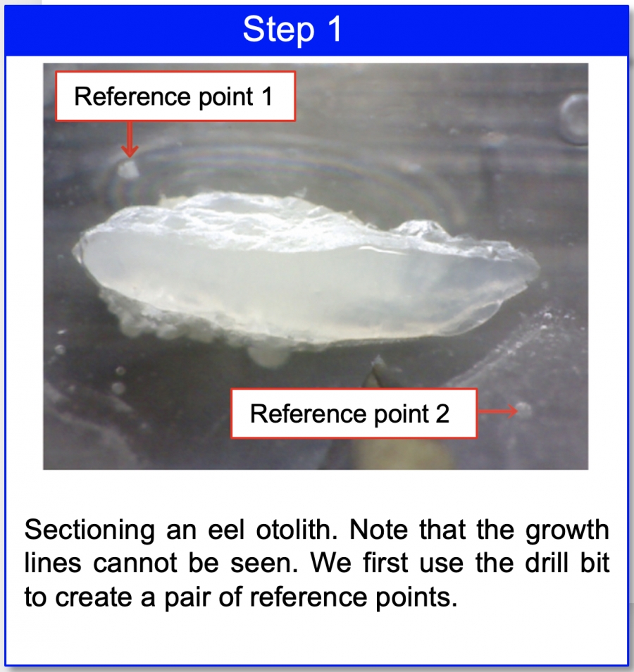
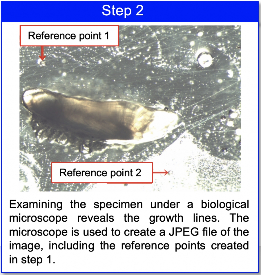
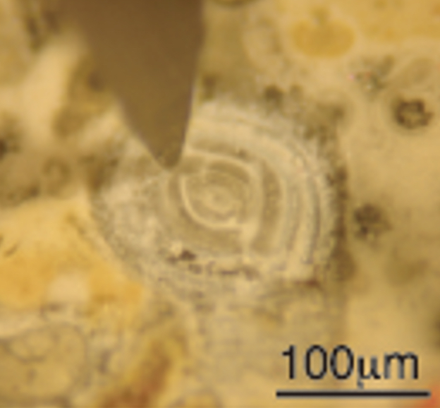
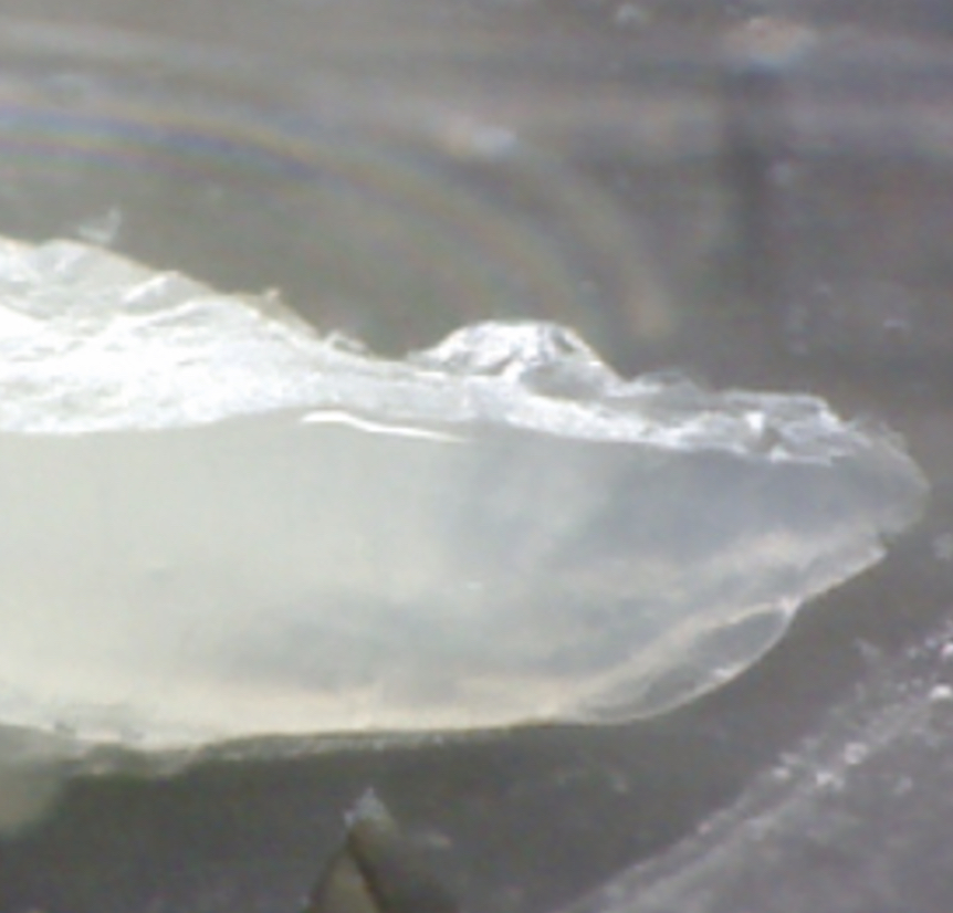
The images below show an example of sectioning an eel otolith. The photo in step 1 is the image as captured by Geomill326, and does not allow observation of the growth lines. As shown in step 2, however, using a high-luminescence biological microscope allows for easy observation of the growth lines.
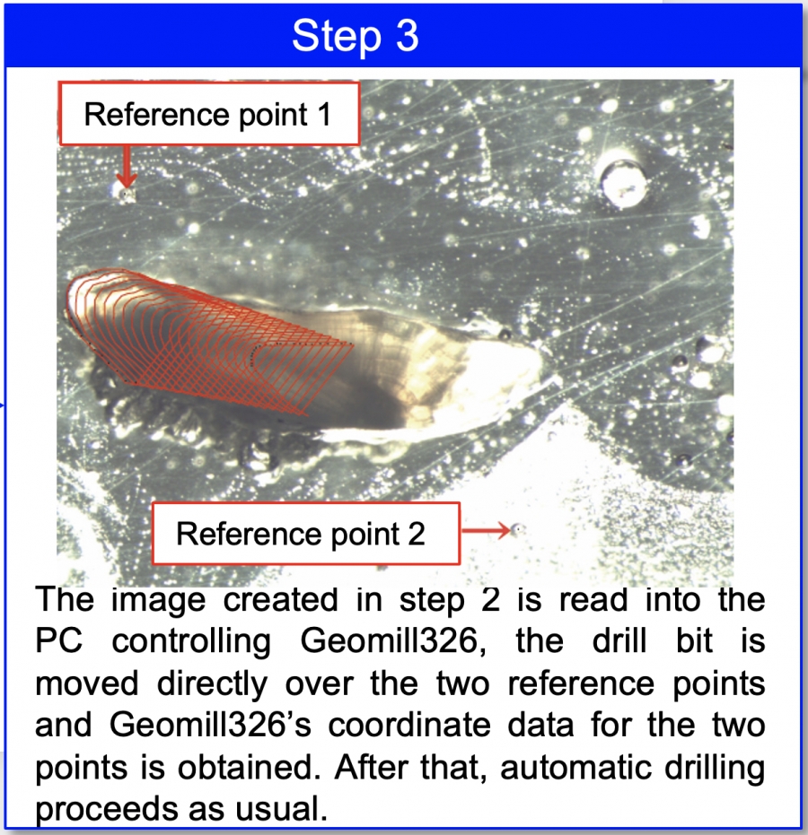
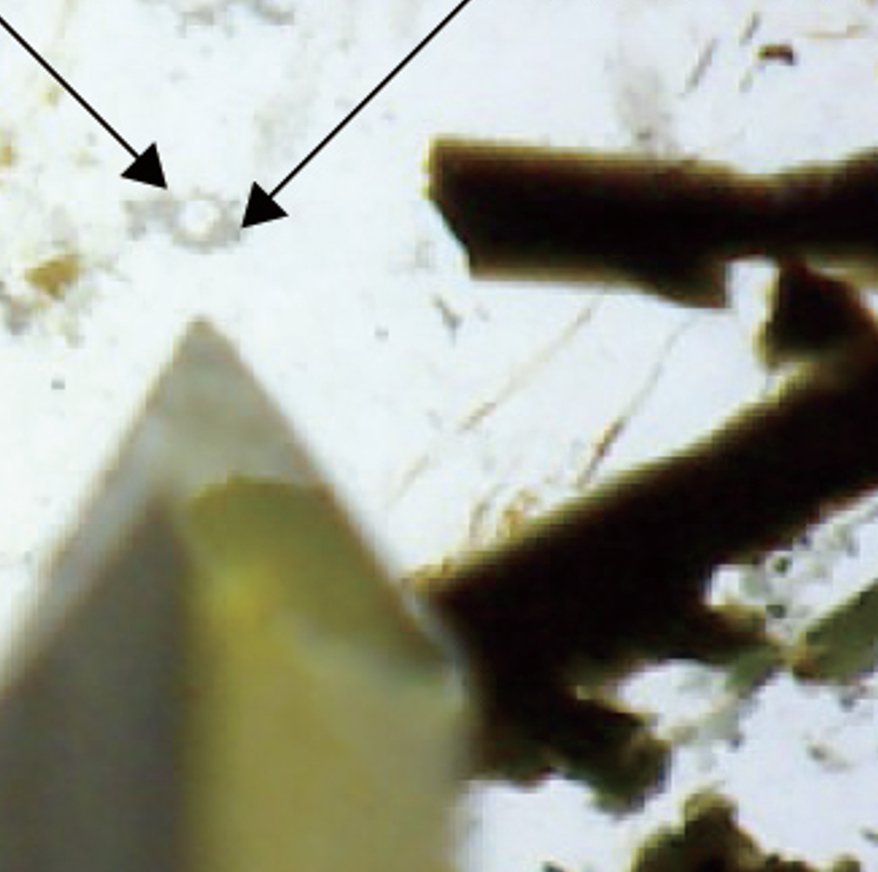
In situations such as this, as shown in step 3 a JPEG file can be saved from the biological microscope image and used as a basis for operating Geomill326.
This is realized by using two reference points added to the drilling specimen on the automatic stage and two corresponding reference points on pixels in the image of the drilling specimen. Together, these can be used to guide drilling.
Example: Use cathodoluminescence images, etc. to cut along zonal structures
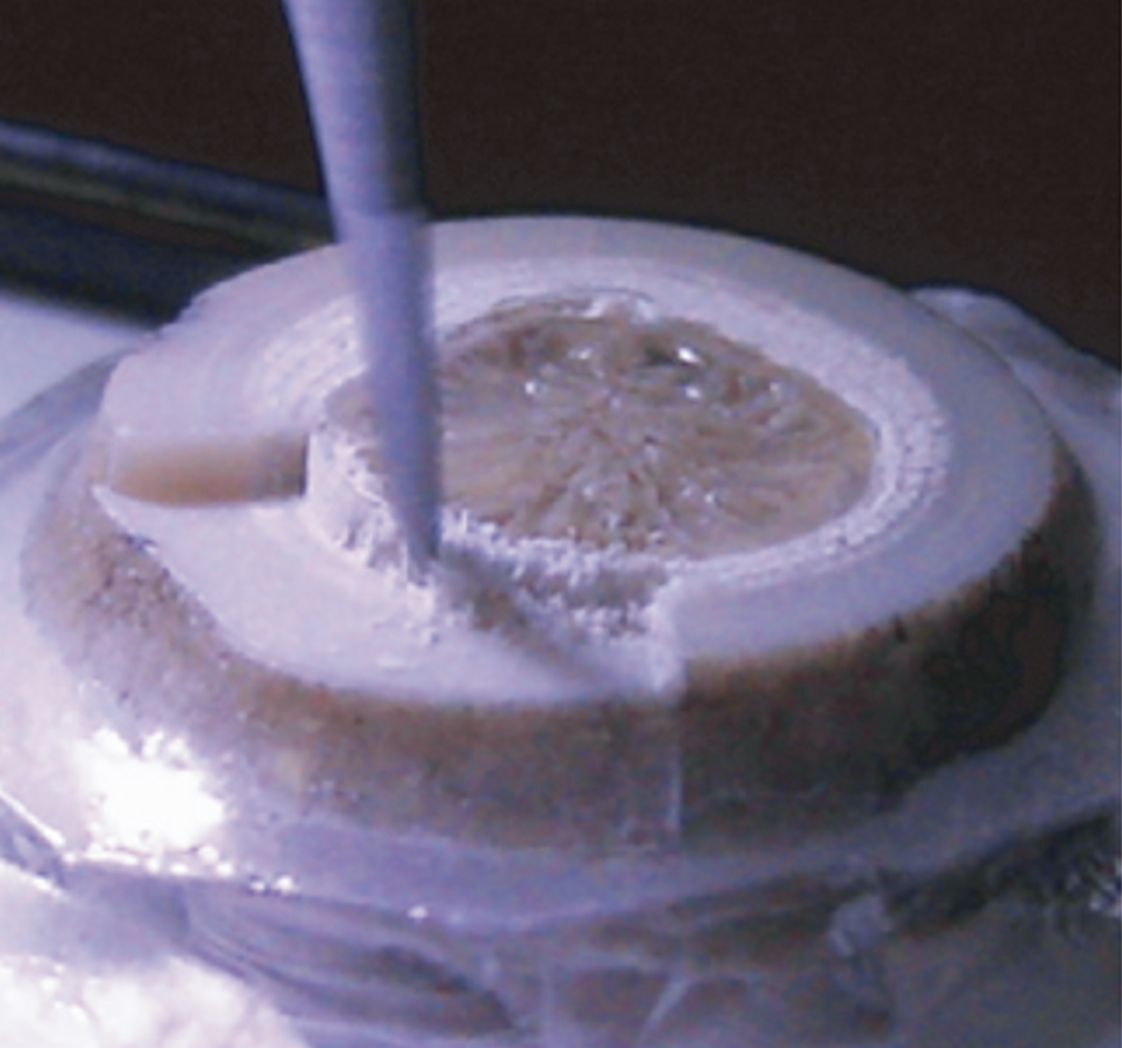
Preparing an appropriate JPEG file containing a pair of reference points can thus allow operation of Geomill326 even on otherwise invisible structures.